Sunday, February 12, 2012
Intrinsic Versus Extrinsic Atopic Dermatitis
Over the past few decades, researchers and clinicians have discovered that there are two forms of AD: intrinsic (nonallergic) and extrinsic
(allergic). Although the two forms are clinically indistinguishable
based on the findings of the physical examination, there are numerous
differences in other aspects (see Table 1 ). Extrinsic AD composes 70%
to 85% of atopic dermatitis, while intrinsic atopic dermatitis makes up
the remaining 15% to 30%. Extrinsic AD is associated with high serum IgE
levels, exhibits allergen-specific IgE to aeroallergens and foods,
positive skin prick reactions, and has a cytokine profile of high IL-4
and IL-13 levels (Bardana, 2004). IL-4, IL-5, and IL-13 cytokines play a
role in the Th2 response which is seen in the early stages of AD.
Chronic atopic dermatitis lesions principally have a Th1 response with
the cytokines IL-12 and interferon-playing a dominant role
(Schmid-Grendelmeier, Simon, Simon, Akdis, & Wuthrich, 2001).
Patients with intrinsic AD, by contrast, have normal IgE levels,
negative skin prick reactions, low IL-4 and IL-13 levels, and they do
not have allergen-specific IgE to aeroallergens and foods (Bardana,
2004). The onset of extrinsic AD typically is in early childhood while
patients with intrinsic AD have a later onset. Finally, patients with
intrinsic AD are characterized by an absence of other atopic disease,
asthma, and allergic rhinitis (Schmid-Grendel meier et al., 2001).
Saturday, February 11, 2012
toxic epidermal necrolysis (TEN)
Patients with toxic epidermal necrolysis (TEN) may describe an influenzalike prodrome characterized by the following:
The cutaneous eruption begins as a poorly defined, erythematous macular rash with purpuric centers. Over a period of hours to days, the rash coalesces to form flaccid blisters and sheetlike epidermal detachment. The lesions predominate on the torso and face, sparing the scalp. Pain at the site of the skin lesions is often the predominating symptom and is often out of proportion to physical findings in early disease.
Mucous membrane erosions (seen in 90% of cases) generally precede the skin lesions by 1-3 days. The most frequently affected mucosal membrane is the oropharynx, followed by the eyes and genitalia. Oral cavity involvement typically presents as a sore or burning sensation. Intake may be limited because of pain associated with the oropharyngeal lesions.
Ocular manifestations range from acute conjunctivitis to corneal erosions and ulcers. Genital involvement may result in painful urination. Other mucosal surfaces such as the esophagus, intestinal tract, or respiratory epithelium may be affected. Bronchial epithelial sloughing may result in dyspnea and hypoxemia.
Most cases of TEN are drug induced, typically occurring within 1-3 weeks of therapy initiation and rarely occurring after more than 8 weeks. Therefore, a detailed medication history, focusing on medications that have been recently started, is a vital component of the patient's history.
More than 220 different medications have been suggested. The most commonly implicated agents include the following:
- Malaise
- Rash
- Fever
- Cough
- Arthralgia
- Myalgia
- Rhinitis
- Headache
- Anorexia
- Nausea and vomiting, with or without diarrhea
The cutaneous eruption begins as a poorly defined, erythematous macular rash with purpuric centers. Over a period of hours to days, the rash coalesces to form flaccid blisters and sheetlike epidermal detachment. The lesions predominate on the torso and face, sparing the scalp. Pain at the site of the skin lesions is often the predominating symptom and is often out of proportion to physical findings in early disease.
Mucous membrane erosions (seen in 90% of cases) generally precede the skin lesions by 1-3 days. The most frequently affected mucosal membrane is the oropharynx, followed by the eyes and genitalia. Oral cavity involvement typically presents as a sore or burning sensation. Intake may be limited because of pain associated with the oropharyngeal lesions.
Ocular manifestations range from acute conjunctivitis to corneal erosions and ulcers. Genital involvement may result in painful urination. Other mucosal surfaces such as the esophagus, intestinal tract, or respiratory epithelium may be affected. Bronchial epithelial sloughing may result in dyspnea and hypoxemia.
Most cases of TEN are drug induced, typically occurring within 1-3 weeks of therapy initiation and rarely occurring after more than 8 weeks. Therefore, a detailed medication history, focusing on medications that have been recently started, is a vital component of the patient's history.
More than 220 different medications have been suggested. The most commonly implicated agents include the following:
- Sulfonamide antibiotics
- Antiepileptic drugs
- Oxicam nonsteroidal anti-inflammatory drugs
- Allopurinol
- Nevirapine
- Abacavir
- Lamotrigine
Physical Examination
Vital signs in toxic epidermal necrolysis may include hyperpyrexia, hypotension secondary to hypovolemia, and tachycardia.
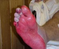 Epidermal sloughing in toxic epidermal necrolysis (TEN).
 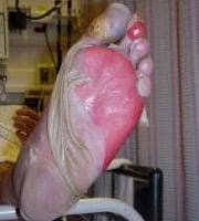 Toxic epidermal necrolysis (TEN) ulcer in great toe (initial infection). A
positive Nikolsky sign is evident when the application of slight
lateral pressure to the epidermal surface results in the epidermis
easily separating from its underlying surface.
Toxic epidermal necrolysis (TEN) ulcer in great toe (initial infection). A
positive Nikolsky sign is evident when the application of slight
lateral pressure to the epidermal surface results in the epidermis
easily separating from its underlying surface.
The usual course is an intense erythema that progresses rapidly to epidermolysis and stops within 2-3 days. Dermatologic recovery typically takes 1-3 weeks, with mucosal lesions taking longer. Rarely, necrolysis recurs in areas that began to heal.
Involvement of the oral mucosa results in edema and erythema, followed by blistering. Ruptured blisters may form extensive hemorrhagic erosions with grayish white pseudomembranes or shallow aphthouslike ulcers. Hemorrhagic crusting of the lips is a common finding (as seen in the image below).
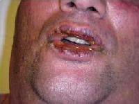 Hemorrhagic crusting of mucous membranes in toxic epidermal necrolysis (TEN). Ocular
involvement varies in severity and can result in mild inflammation,
conjunctival erosion, purulent exudates, or pseudomembrane formation.
Hemorrhagic crusting of mucous membranes in toxic epidermal necrolysis (TEN). Ocular
involvement varies in severity and can result in mild inflammation,
conjunctival erosion, purulent exudates, or pseudomembrane formation.
Involvement of respiratory epithelium may result in bronchial hypersecretion, hypoxemia, interstitial infiltrates, pulmonary edema, bacterial pneumonia, or bronchiolitis obliterans.
Skin examination
Skin lesions begin as painful/burning, warm, erythematous, morbilliform macules that are initially discrete. They begin symmetrically on the face and thorax before spreading to the entire body. The skin lesions coalesce and fill with fluid-producing large, flaccid blisters. The epidermis sloughs in sheets, leaving a characteristic moist, denuded dermis (see images below). Conjunctivitis and denudation and erosions of other mucous membranes precede epidermal necrolysis.Epidermal sloughing in toxic epidermal necrolysis (TEN). Toxic epidermal necrolysis (TEN) ulcer in great toe (initial infection). A
positive Nikolsky sign is evident when the application of slight
lateral pressure to the epidermal surface results in the epidermis
easily separating from its underlying surface. The usual course is an intense erythema that progresses rapidly to epidermolysis and stops within 2-3 days. Dermatologic recovery typically takes 1-3 weeks, with mucosal lesions taking longer. Rarely, necrolysis recurs in areas that began to heal.
Involvement of the oral mucosa results in edema and erythema, followed by blistering. Ruptured blisters may form extensive hemorrhagic erosions with grayish white pseudomembranes or shallow aphthouslike ulcers. Hemorrhagic crusting of the lips is a common finding (as seen in the image below).
Hemorrhagic crusting of mucous membranes in toxic epidermal necrolysis (TEN). Ocular
involvement varies in severity and can result in mild inflammation,
conjunctival erosion, purulent exudates, or pseudomembrane formation. Involvement of respiratory epithelium may result in bronchial hypersecretion, hypoxemia, interstitial infiltrates, pulmonary edema, bacterial pneumonia, or bronchiolitis obliterans.
Complications
Numerous
complications of TEN can arise as a result of the widespread cutaneous
and mucosal membrane inflammation and necrosis. Stomatitis and
mucositis, which are painful and hinder oral intake, can place patients
at risk for dehydration and malnutrition.
Epithelial loss predisposes to septicemia (Pseudomonas aeruginosa, Staphylococcus aureus, gram-negative species, and Candida albicans). Renal hypoperfusion, acute tubular necrosis, and renal insufficiency may develop subsequent to septic shock.
Ulceration of various mucosal membranes may result in pain, scarring, and stricture formation. Affected surfaces include oral, ocular, and urogenital mucosa. Barrera and colleagues reported a case of hypopharyngeal stenosis and dysphagia with recurrent aspiration.[27] Miscellaneous complications include hypovolemia, massive gut bleeding, and pulmonary emboli.
Mild-to-severe eye complications can occur, such as the following:
GI hemorrhage results from intestinal inflammation.
Epithelial loss predisposes to septicemia (Pseudomonas aeruginosa, Staphylococcus aureus, gram-negative species, and Candida albicans). Renal hypoperfusion, acute tubular necrosis, and renal insufficiency may develop subsequent to septic shock.
Ulceration of various mucosal membranes may result in pain, scarring, and stricture formation. Affected surfaces include oral, ocular, and urogenital mucosa. Barrera and colleagues reported a case of hypopharyngeal stenosis and dysphagia with recurrent aspiration.[27] Miscellaneous complications include hypovolemia, massive gut bleeding, and pulmonary emboli.
Mild-to-severe eye complications can occur, such as the following:
- Lid edema
- Persistent dry eyes
- Chronic photosensitivity
- Conjunctivitis
- Keratitis
- Conjunctival fornix foreshortening
- Symblepharon
- Corneal ulceration and scarring
- Blindness
GI hemorrhage results from intestinal inflammation.
Staphylococcal Scalded Skin Syndrome
Staphylococcal scalded skin syndrome (SSSS) presents as a red rash followed by diffuse epidermal exfoliation.
A prodromal localized S aureus infection of the skin, throat, nose, mouth, umbilicus, or GI tract occurs. Such an infection often is not apparent before the SSSS rash appears.
The following may be noted:
A prodromal localized S aureus infection of the skin, throat, nose, mouth, umbilicus, or GI tract occurs. Such an infection often is not apparent before the SSSS rash appears.
The following may be noted:
- General malaise
- Fever
- Irritability
- Skin tenderness
Physical
The following may be noted:- Fever, although patients may be afebrile
- Tenderness to palpation
- Warmth to palpation
- Facial edema
- Perioral crusting
- Most patients do not appear severely ill.
- Dehydration may be present and significant.
- Nikolsky sign (gentle stroking of the skin causes the skin to separate at the epidermis)[20, 21]
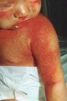 Staphylococcal scalded skin syndrome. Photograph by David Effron, MD, FACEP.
  Staphylococcal scalded skin syndrome. Photograph by David Effron, MD, FACEP. Bullae are flaccid and ill defined. See the image below.
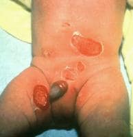 Staphylococcal scalded skin syndrome. Photograph by David Effron, MD, FACEP. Exfoliation of skin, which may be patchy or sheetlike in nature, is noted. See the images below.
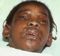 Staphylococcal scalded skin syndrome. Photograph by David Effron, MD, FACEP
cutaneous larva migrans
- Tingling/prickling at the site of exposure within 30 minutes of penetration of larvae, although Archer describes a case of late-onset cutaneous larva migrans (CLM)[6]
- Intense pruritus
- Erythematous, often linear lesions that advance
- Often associated with a history of sunbathing, walking barefoot on the beach, or similar activity in a tropical location
- Predispositions to contracting cutaneous larva migrans include the following:
- Hobbies and occupations that involve contact with warm, moist, sandy soil
- Tropical/subtropical climate travel
- Barefoot beachgoers/sunbathers
- Children in sandboxes
- Carpenter
- Electrician
- Plumber
- Farmer
- Gardener
- Pest exterminator
Physical
- Cutaneous signs of cutaneous larva migrans (CLM) include the following:
- Pruritic, erythematous, edematous papules and/or vesicles
- Serpiginous (snakelike), slightly elevated, erythematous tunnels that are 2- to 3-mm wide and track 3-4 cm from the penetration site
- Nonspecific dermatitis
- Vesicles with serous fluid
- Secondary impetiginization
- Tract advancement of 1-2 cm/d
- Systemic signs include peripheral eosinophilia (Loeffler syndrome), migratory pulmonary infiltrates, and increased immunoglobulin E (IgE) levels, but are rarely seen.
- Lesions are typically distributed on the distal lower extremities, including the dorsa of the feet and the interdigital spaces of the toes, but can also occur in the anogenital region, the buttocks, the hands, and the knees


Subscribe to:
Comments (Atom)