Nummular
(meaning "coin-shaped") dermatitis is a form of eczema. Nummular
dermatitis is characterized by round-to-oval erythematous plaques most
commonly found on the arms and legs. Lesions often start as papules,
which then coalesce into plaques with scale. Early nummular dermatitis
lesions may be studded with vesicles containing serous exudate. They are
usually very pruritic.
Pathophysiology
Nummular dermatitis is a condition confined to the skin. It has recently been classified as a form of atopic dermatitis.
Little is known about the pathophysiology of nummular dermatitis, but it is frequently accompanied by xerosis. Dryness of the skin results in dysfunction of the epidermal lipid barrier; this may allow permeation of environmental allergens, which induce an allergic or irritant response. This is supported by one study that showed that elderly patients with nummular dermatitis had increased sensitivity to environmental aeroallergens compared with age-matched controls. This impaired cutaneous barrier in the setting of nummular dermatitis may also lead to increased susceptibility to allergic contact dermatitis to materials such as metals.
Onset has been associated with medications. Onset of severe, generalized nummular lesions has been reported in association with interferon and ribavirin therapy for hepatitis C. Association with use of inhibitors of tumor necrosis factor has also been reported.
Onset has also been described in association with mercury in dental amalgams. Hypersensitivity to the metals in the mouth is posulated to be sufficient to drive an immune response that results in cutaneous nummular plaques.
Because of the intense pruritus associated with nummular dermatitis, the potential role of mast cells in the disease process has been investigated. Increased numbers of mast cells have been observed in lesional compared with nonlesional samples in persons with nummular dermatitis.
One study identified neurogenic contributors to inflammation in both nummular dermatitis and atopic dermatitis by investigating the association between mast cells and sensory nerves and identifying the distribution of neuropeptides in the epidermis and upper dermis of patients with nummular eczema. Researchers hypothesized that release of histamine and other inflammatory mediators from mast cells may initiate pruritus by interacting with neural C-fibers. The research showed that dermal contacts between mast cells and nerves were increased in number in both lesional and nonlesional samples of nummular eczema compared with normal controls. In addition, substance P and calcitonin gene-related peptide fibers were prominently increased in lesional samples compared with nonlesional samples from patients with nummular eczema. These neuropeptides may stimulate release of other cytokines and promote inflammation.[6, 7, 8]
Other research has demonstrated that mast cells present in the dermis of patients with nummular eczema may have decreased chymase activity, imparting reduced ability to degrade neuropeptides and protein. This dysregulation could lead to decreased capability of the enzyme to suppress inflammation.
Epidemiology
Frequency
United States
The prevalence of nummular dermatitis is 2 cases per 1000 people. Dermatitis (eg, atopic, asteatotic, dyshidrotic, nummular, hand) is one of the most common dermatologic conditions.International
The incidence internationally is the same as it is in the United States.Mortality/Morbidity
- Pruritus, often worst at night, may cause irritability, insomnia, or both.
- Secondary infection may result in lesions that ooze serosanguineous exudate. The most common organism revealed by culture is Staphylococcus aureus.
- Generalized flares may require bed rest, oral antibiotics, a cool environment, systemic antibiotics, and/or systemic steroids.
- Increased contact sensitivity to environmental antigens (especially metals) could limit ability to tolerate those antigens, especially clothing, metal snaps, jewelry, dental amalgams or occupational exposure.
Race
No racial predilection has been observed for nummular dermatitis.Sex
Nummular dermatitis is more common in males than in females (see Age below).Age
Nummular dermatitis has 2 peaks of age distribution. The most common is in the sixth to seventh decade of life. This is most often seen in males. A smaller peak occurs in the second to third decade of life, which is most often seen in association with atopic dermatitis. This is more often seen in females. It is uncommon in children.
Patients present with a days-to-months' history of a pruritic eruption, usually starts on the legs. It may also burn or sting.
- Nummular dermatitis often waxes and wanes with winter; cold or dry climates or swings in temperature may be exacerbating factors. It may improve with sun or humidity exposure or with moisturizer use. Occasionally it may worsen with heat or humidity.
- New nummular dermatitis lesions often recur in the same locations as old lesions.
- The patient's medical history may be positive for eczema, atopic dermatitis, or dry and sensitive skin.
Physical
The
diagnosis of nummular dermatitis is made on the basis of observing the
characteristic round-to-oval erythematous plaques. They are most
commonly located on the extremities, particularly the legs, but they may
occur anywhere on the trunk, hands, or feet. Nummular dermatitis does
not involve the face and scalp. Lesions are often symmetrically
distributed.
See the image below.
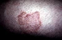 Dry, scaling plaque of nummular dermatitis (size, 3 X 5 cm) on the shin. Lesions
begin as erythematous-to-violaceous papules or vesicles, which then
coalesce to form confluent plaques. They may have overlying erosions due
to excoriation.
Dry, scaling plaque of nummular dermatitis (size, 3 X 5 cm) on the shin. Lesions
begin as erythematous-to-violaceous papules or vesicles, which then
coalesce to form confluent plaques. They may have overlying erosions due
to excoriation.
Early lesions, particularly vesicular ones, often become colonized by staphylococci, which produces a yellowish crust. Secondary overt infection may occur, with cellulitis surrounding the plaques, requiring oral antibiotics.
Within a few days, plaques become dry, scaly, and more violaceous, particularly when located below the knee.
The lesions then flatten to macules, usually with brown postinflammatory hyperpigmentation that gradually lightens. The pigment may never completely fade, particularly when located below the knee.
Plaques may show central clearing, making differentiation from tinea corporis based on clinical findings difficult. Tinea corporis usually has few vesicles, a raised narrow border, and leading scale (ie, scale on the outside of the plaque).
Distinguishing between forms of dermatitis (eg, asteatotic eczema, atopic dermatitis, nummular dermatitis) may be difficult, but, fortunately, this is not necessary to make proper treatment decisions. Contact dermatitis may have a pattern that approximates the manner in which the offending agent came into contact with the skin, such as a linear pattern. It may become chronic in the setting of repeated exposure, such as with chromates and formaldehyde. The patient may recall contact with an allergen, such as poison ivy.
Lichen simplex chronicus often occurs on the lower legs, the neck, the scalp, or the scrotum; it is lichenified (thickened by chronic scratching), more violaceous, and, often, has no clear border.
Stasis dermatitis may occur simultaneously on the lower extremities, and venous stasis may lead to the concomitant development of both conditions.
Psoriasis plaques are often found on the extensor surfaces, especially at the elbows and knees, in addition to other areas. The scalp is often involved. Psoriasis scale is usually thick and silver and bleeds when removed (Auspitz sign).
See the image below.
Dry, scaling plaque of nummular dermatitis (size, 3 X 5 cm) on the shin. Lesions
begin as erythematous-to-violaceous papules or vesicles, which then
coalesce to form confluent plaques. They may have overlying erosions due
to excoriation.Early lesions, particularly vesicular ones, often become colonized by staphylococci, which produces a yellowish crust. Secondary overt infection may occur, with cellulitis surrounding the plaques, requiring oral antibiotics.
Within a few days, plaques become dry, scaly, and more violaceous, particularly when located below the knee.
The lesions then flatten to macules, usually with brown postinflammatory hyperpigmentation that gradually lightens. The pigment may never completely fade, particularly when located below the knee.
Plaques may show central clearing, making differentiation from tinea corporis based on clinical findings difficult. Tinea corporis usually has few vesicles, a raised narrow border, and leading scale (ie, scale on the outside of the plaque).
Distinguishing between forms of dermatitis (eg, asteatotic eczema, atopic dermatitis, nummular dermatitis) may be difficult, but, fortunately, this is not necessary to make proper treatment decisions. Contact dermatitis may have a pattern that approximates the manner in which the offending agent came into contact with the skin, such as a linear pattern. It may become chronic in the setting of repeated exposure, such as with chromates and formaldehyde. The patient may recall contact with an allergen, such as poison ivy.
Lichen simplex chronicus often occurs on the lower legs, the neck, the scalp, or the scrotum; it is lichenified (thickened by chronic scratching), more violaceous, and, often, has no clear border.
Stasis dermatitis may occur simultaneously on the lower extremities, and venous stasis may lead to the concomitant development of both conditions.
Psoriasis plaques are often found on the extensor surfaces, especially at the elbows and knees, in addition to other areas. The scalp is often involved. Psoriasis scale is usually thick and silver and bleeds when removed (Auspitz sign).
Causes
The etiology is unknown and likely multifactorial.Most patients with nummular dermatitis also have very dry (xerotic) skin.
Local trauma, such as arthropod bites, contact with chemicals, or abrasions, may precede an outbreak.
Contact dermatitis may play a role in some cases. Contact dermatitis may be irritant or allergic in nature. Sensitivity to nickel, cobalt, or chromates has been reported in patients with nummular dermatitis. In one study, the most frequent sensitizers were colophony, nitrofurazone, neomycin sulfate, and nickel sulfate. In the past, cases of nummular eczema–like eruptions have been caused by ethyl cyanoacrylate–containing glue, thimerosal, mercury-containing dental amalgams, and depilating creams containing potassium thioglycolate.
Venous insufficiency (and varicosities), stasis dermatitis, and edema may be related to involvement of the affected lower extremities.
Autoeczematization (ie, lesional spread from the initial focal site) may account for the presence of multiple plaques.
Onset of severe, generalized nummular lesions has been reported in association with interferon therapy for hepatitis C as well as exposure to mercury.
Various types of eczematous eruptions, including nummular dermatitis, have been observed following tumor necrosis factor-alpha blocking therapy.
Nummular eczema has been found in association with infection in rare cases. Giardiasis has been reported. One study reported that in patients with Helicobacter pylori infection and nummular dermatitis, eradication of H pylori caused clearance of the skin lesions in 54% of the patients. Another case report noted nummular eczema in association with a dental infection that cleared after the treatment of the infection
Treatment is aimed at rehydration of the skin and repair of the
epidermal lipid barrier, reduction of inflammation and treatment of any
infection.
Lukewarm or cool baths or showers reduce itching and help rehydrate the skin. Patients should be instructed to bathe 1-2 times a day at least, followed by the application of moisturizers or medicated topical preparations to seal the water in the skin. The "soak-and-smear" therapeutic regimen includes a 20-minute plain water soak each night followed by application of steroid ointment or petrolatum to wet skin and includes alteration of cleansing habits so that soap is applied only to the axilla and groin. One study showed greater than 90% response in 27 of 28 patients with refractory chronic pruritic eruptions when the regimen was followed as directed.
Wet wrap treatments are often helpful. This involves dampening the skin with lukewarm water until it is well hydrated (usually 10 min). Then, petrolatum or steroid ointment is applied liberally, followed by occlusion for 1 hour with damp pajamas or a nonbreathable sauna suit. For small areas of involvement, plastic wrap may be used to occlude the area. This process may be repeated 5-6 times a day with petrolatum. Caution must be used when prescription steroid medications are used because overuse of these medications can cause striae, thinning of the skin, and, rarely, enough systemic absorption of steroid to affect the hypothalamic-pituitary-adrenal axis.
Steroids are the most commonly used therapy to reduce inflammation.
Topical steroids are effective. Less erythematous, less pruritic lesions may be treated with low-potency (class III-VI) steroids. Severely inflamed lesions with intense erythema, vesicles, and pruritus require high-potency (class I-II) preparations. Penetration of the medication is enhanced by occlusion or presoaking in a tub of plain water followed immediately (without drying) by application of the steroid-containing ointment.
Application of the medicine to damp skin allows more effective penetration and faster healing.
Ointments are usually more effective than creams because they are more occlusive, form a barrier between the skin and the environment, and more effectively hold water into the skin.
Emollients and topical class I-III topical steroids may be used short term.
Oral, intramuscular, or parenteral steroids may be required in cases of severe, generalized eruptions.
Tar preparations are helpful to decrease inflammation, particularly in older, thickened, scaly plaques.
Topical immune modulators (tacrolimus and pimecrolimus) also reduce inflammation. These are often initiated a few days after the topical steroid to decrease the risk of a burning sensation that may occur when applied to extremely irritated skin.
When eruptions are generalized and prolonged, phototherapy (generally UVB) may be helpful.
Oral antihistamines or sedatives may help reduce itching and improve sleep.
Oral antibiotics, such as dicloxacillin, cephalexin, or erythromycin, should be used in cases of secondary infection. Swab cultures of the skin guide selection of antibiotics.
Phototherapy may be helpful. Broadband or narrow band UVB is most commonly used, although PUVA (Psoralen + UVA) may be used in severe cases.
Once the eruption has resolved, ongoing aggressive hydration may decrease the frequency between flares, particularly in dry climates. Heavy moisturizers (preferably a sensitive-skin formulation) or petroleum jelly applied to damp skin after showering may be helpful.
Disease may be severe and refractory to the above treatments. Immune suppressive medications such as methotrexate have been described to be safe and effective in these severly affected patients.
nice blog thanks for commenting
ReplyDeleteyour tips are very useful. thanks for posting.
skin care clinic
ReplyDeleteBut Dr. Itua, Traditional Herbal Practitioner in Africa, Have cured for HIV which is extracted from some rare herbals. It is highly potential to cure AIDS 100% without any residue. Dr Itua herbal medicine has already passed various blogs on how he use his powerful herbals to heal all kind of diseases such as. Herpes, HIV,,Cushing’s disease,Heart failure,Multiple Sclerosis,Hypertension,Colo_Rectal Cancer, Diabetes, Hepatitis,Hpv,Weak ErectionLyme Disease,Blood Cancer,Alzheimer’s disease,Bechet’s disease,Crohn’s disease,Parkinson's disease,Schizophrenia,Lung Cancer,Breast Cancer,Colo-Rectal Cancer,Blood Cancer,Prostate Cancer,siva.Fatal Familial Insomnia Factor V Leiden Mutation ,Epilepsy Dupuytren's disease,Desmoplastic small-round-cell tumor Diabetes ,Coeliac disease,Creutzfeldt–Jakob disease,Cerebral Amyloid Angiopathy, Ataxia,Arthritis,Amyotrophic Lateral Scoliosis,Fibromyalgia,Fluoroquinolone Toxicity,Brain Cancer,Breast Cancer,Lung Cancer,Kidney Cancer,Syndrome Fibrodysplasia Ossificans ProgresSclerosis,Seizures,Alzheimer's disease,Adrenocortical carcinoma.Asthma,Allergic diseases.Hiv_ Aids,Herpe ,Copd,Glaucoma., Cataracts,Macular degeneration,Cardiovascular disease,Lung disease.Enlarged prostate,Osteoporosis.Alzheimer's disease,
Dementia.,Wart Remover,Cold Sore, Epilepsy, also his herbal boost immune system as well. I'm telling this because he uses his herbal medicine to cure me from hepatitis B and HIV, which i have being living for 9 months now with no side effect. The Herbal Medicine is just as good when drinking it although i have to use rest room after drinking it which I do not really care about because i just want to get the virus out of my body, I will recommend Dr Itua to anyone sick out here to contact Dr Itua with this following information.
Email...drituaherbalcenter@gmail.com /
Whatsapp Or Call...+2348149277967.
He might be late to respond because he always busy with patent, but he will surely get back to you with positive response.