Pemphigus is derived from the Greek word pemphix
meaning bubble or blister. Pemphigus describes a group of chronic
bullous diseases, originally named by Wichman in 1791. The term
pemphigus once included most bullous eruptions of the skin, but
diagnostic tests have improved, and bullous diseases have been
reclassified.
The term pemphigus refers to a group of autoimmune blistering diseases of the skin and mucous membranes characterized histologically by intraepidermal blister and immunopathologically by the finding of in vivo bound and circulating immunoglobulin G (IgG) antibody directed against the cell surface of keratinocytes. The 3 primary subsets of pemphigus include pemphigus vulgaris, pemphigus foliaceus, and paraneoplastic pemphigus. Each type of pemphigus has distinct clinical and immunopathologic features. Pemphigus vulgaris accounts for approximately 70% of pemphigus cases.
The term pemphigus refers to a group of autoimmune blistering diseases of the skin and mucous membranes characterized histologically by intraepidermal blister and immunopathologically by the finding of in vivo bound and circulating immunoglobulin G (IgG) antibody directed against the cell surface of keratinocytes. The 3 primary subsets of pemphigus include pemphigus vulgaris, pemphigus foliaceus, and paraneoplastic pemphigus. Each type of pemphigus has distinct clinical and immunopathologic features. Pemphigus vulgaris accounts for approximately 70% of pemphigus cases.
Pathophysiology
Pemphigus
vulgaris is an autoimmune, intraepithelial, blistering disease
affecting the skin and mucous membranes and is mediated by circulating
autoantibodies directed against keratinocyte cell surfaces. In 1964,
autoantibodies against keratinocyte surfaces were described in patients
with pemphigus. Clinical and experimental observations indicate that the
circulating autoantibodies are pathogenic. An immunogenetic
predisposition is well established.
Blisters in pemphigus vulgaris are associated with the binding of IgG autoantibodies to keratinocyte cell surface molecules. These intercellular or pemphigus vulgaris antibodies bind to keratinocyte desmosomes and to desmosome-free areas of the keratinocyte cell membrane. The binding of autoantibodies results in a loss of cell-to-cell adhesion, a process termed acantholysis. The antibody alone is capable of causing blistering without complement or inflammatory cells.
More than 80% of the patients with active disease produce autoantibodies to the desmosomal protein desmoglein. Disease activity correlates with antibody titers in most patients. In patients with pemphigus vulgaris, the presence of antidesmoglein 1 autoantibodies, as determined by enzyme-linked immunosorbent assay (ELISA), is more closely correlated with the course of the disease compared with antidesmoglein 3 autoantibodies. Lack of in vivo antibody binding (reversion to a negative result on direct immunofluorescence) is the best indicator of remission and can help predict a lack of flaring when therapy is tapered.
Blisters in pemphigus vulgaris are associated with the binding of IgG autoantibodies to keratinocyte cell surface molecules. These intercellular or pemphigus vulgaris antibodies bind to keratinocyte desmosomes and to desmosome-free areas of the keratinocyte cell membrane. The binding of autoantibodies results in a loss of cell-to-cell adhesion, a process termed acantholysis. The antibody alone is capable of causing blistering without complement or inflammatory cells.
Pemphigus vulgaris antigen
Intercellular adhesion in the epidermis involves several keratinocyte cell surface molecules. Pemphigus antibody binds to keratinocyte cell surface the molecules desmoglein 1 and desmoglein 3. The binding of antibody to desmoglein may have a direct effect on desmosomal adherens or may trigger a cellular process that results in acantholysis. Antibodies specific for nondesmosomal antigens also have been described in the sera of patients with pemphigus vulgaris; however, the role of these antigens in the pathogenesis of pemphigus vulgaris is not known.Antibodies
Patients with the mucocutaneous form of pemphigus vulgaris have pathogenic antidesmoglein 1 and antidesmoglein 3 autoantibodies. Patients with the mucosal form of pemphigus vulgaris have only antidesmoglein 3 autoantibodies. Patients with active disease have circulating and tissue-bound autoantibodies of both the immunoglobulin G1 (IgG1) and immunoglobulin G4 (IgG4) subclasses.More than 80% of the patients with active disease produce autoantibodies to the desmosomal protein desmoglein. Disease activity correlates with antibody titers in most patients. In patients with pemphigus vulgaris, the presence of antidesmoglein 1 autoantibodies, as determined by enzyme-linked immunosorbent assay (ELISA), is more closely correlated with the course of the disease compared with antidesmoglein 3 autoantibodies. Lack of in vivo antibody binding (reversion to a negative result on direct immunofluorescence) is the best indicator of remission and can help predict a lack of flaring when therapy is tapered.
Complement
Pemphigus antibody fixes components of complement to the surface of epidermal cells. Antibody binding may activate complement with the release of inflammatory mediators and recruitment of activated T cells. T cells are clearly required for the production of the autoantibodies, but their role in the pathogenesis of pemphigus vulgaris remains poorly understood. Interleukin 2 is the main activator of T lymphocytes, and increased soluble receptors have been detected in patients with active pemphigus vulgaris.Epidemiology
Frequency
United States
Pemphigus vulgaris is uncommon, and the exact incidence and prevalence depends on the population studied.International
Pemphigus vulgaris has been reported to occur worldwide. Pemphigus vulgaris incidence varies from 0.5-3.2 cases per 100,000 population. Pemphigus vulgaris incidence is increased in patients of Ashkenazi Jewish descent and those of Mediterranean origin. Few familial cases have been reported.Mortality/Morbidity
Pemphigus vulgaris is a potentially life-threatening autoimmune mucocutaneous disease with a mortality rate of approximately 5-15%. Mortality in patients with pemphigus vulgaris is 3 times higher than the general population. Complications secondary to the use of high-dose corticosteroids contribute to the mortality rate. Morbidity and mortality are related to the extent of disease, the maximum dose of systemic steroids required to induce remission, and the presence of other diseases. Prognosis is worse in patients with extensive pemphigus vulgaris and in older patients.Pemphigus vulgaris involves mucosa in 50-70% of patients. This may limit oral intake secondary to dysphagia. Blistering and erosions secondary to the rupture of blisters may be painful and may limit the patient's daily activities. Additionally, Patients with pemphigus vulgaris typically heal without scarring unless the disease is complicated by severe secondary infection.
Race
Pemphigus vulgaris affects persons of all races. The prevalence of pemphigus vulgaris is high in regions where the Jewish population is predominant.[6] For example, in Jerusalem, the prevalence of pemphigus vulgaris is estimated at 1.6 cases per 100,000 population; in Connecticut, the prevalence has been reported as 0.42 cases per 100,000 population. The incidence in the United Kingdom is 0.68 case per 100 000 persons per year. The incidence of pemphigus vulgaris in Tunisia is estimated at 2.5 cases per million population per year (3.9 in women, 1.2 in men), while in France, the incidence is 1.3 cases per million population per year (no significant difference between men and women) In Finland, where few people of Jewish or Mediterranean origin live, the prevalence is low, at 0.76 case per million population.Sex
The male-to-female ratio is approximately equal. In adolescence, girls are more likely to be affected than boys.Age
The mean age of onset is approximately 50-60 years; however, the range is broad, and disease onset in older individuals and in children has been described. Patients are younger at presentation in India than in Western countries.- Mucous membranes: Pemphigus vulgaris presents with oral lesions in 50-70% of patients, and almost all patients have mucosal lesions at some point in the course of their disease. Mucosal lesions may be the sole sign for an average of 5 months before skin lesions develop, or they may be the sole manifestation of the disease. The diagnosis of pemphigus vulgaris should be considered in any patient with persistent oral erosive lesions.
- Skin: Most patients with pemphigus vulgaris develop cutaneous lesions. The primary lesion of pemphigus vulgaris is a flaccid blister, which usually arises on healthy-appearing skin but may be found on erythematous skin. New blisters usually are flaccid or become flaccid quickly. Affected skin often is painful but rarely pruritic.
- Drug-induced pemphigus vulgaris : Drugs reported most significantly in association with pemphigus vulgaris include penicillamine, captopril, cephalosporin, pyrazolones, nonsteroidal anti-inflammatory drugs (NSAIDs), and other thiol-containing compounds. Rifampin, emotional stress, thermal burns, ultraviolet rays, and infections (eg, coxsackievirus, Herpesviridae family) have also been reported as triggers for pemphigus vulgaris.
Physical
Mucous
membranes typically are affected first in pemphigus vulgaris. Mucosal
lesions may precede cutaneous lesions by weeks or months. Patients with
mucosal lesions may present to dentists, oral surgeons, or
gynecologists.[13]
- Mucous membranes
- Intact bullae are rare in the mouth. More commonly, patients have ill-defined, irregularly shaped, gingival, buccal, or palatine erosions, which are painful and slow to heal. The erosions extend peripherally with shedding of the epithelium.
- The mucous membranes most often affected in pemphigus vulgaris are those of the oral cavity, which is involved in almost all patients with pemphigus vulgaris and sometimes is the only area involved. Erosions may be seen on any part of the oral cavity. Erosions can be scattered and often are extensive. Erosions may spread to involve the larynx, with subsequent hoarseness. The patient often is unable to eat or drink adequately because the erosions are so uncomfortable.
- In juvenile pemphigus vulgaris, stomatitis is the presenting complaint in more than 50% of the cases.
- Other mucosal surfaces may be involved, including the conjunctiva, esophagus (causes odynophagia and/or dysphagia), labia, vagina, cervix, vulva, penis, urethra, nasal mucosa, and anus.
- Skin
- The
primary lesion of pemphigus vulgaris is a flaccid blister filled with
clear fluid that arises on healthy skin or on an erythematous base, as
shown in the images below.
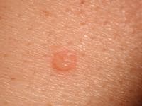 Early, small blister filled with clear fluid arises on healthy skin.
 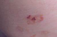 Flaccid blister filled with clear fluid arises on healthy skin.
 - The
blisters are fragile; therefore, intact blisters may be sparse. The
contents soon become turbid, or the blisters rupture, producing painful
erosions, which is the most common skin presentation and is shown in the
image below. Erosions often are large because of their tendency to
extend peripherally with the shedding of the epithelium.
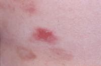 An erosion.
- The
primary lesion of pemphigus vulgaris is a flaccid blister filled with
clear fluid that arises on healthy skin or on an erythematous base, as
shown in the images below.
- Vegetating pemphigus vulgaris: Ordinary pemphigus vulgaris erosions may develop vegetation. Lesions in skin folds readily form vegetating granulations. In some patients, erosions tend to develop excessive granulation tissue and crusting, and these patients display more vegetating lesions. This type of lesion tends to occur more frequently in intertriginous areas and on the scalp or face. The vegetating type of response can be more resistant to therapy and can remain in one place for long periods.
- Pemphigus in pregnancy: Pemphigus vulgaris occurring in pregnancy is rare. When present, maternal autoantibodies may cross the placenta, resulting in neonatal pemphigus. Neonatal pemphigus is transient and improves with clearance of maternal autoantibodies.Treatment of pemphigus vulgaris in pregnancy is with oral corticosteroids; however, prednisone and its metabolites cross the placenta and have been associated with low birth weight, prematurity, infection, and adrenal insufficiency.
- Nikolsky sign: In patients with active blistering, firm sliding pressure with a finger separates normal-appearing epidermis, producing an erosion. This sign is not specific for pemphigus vulgaris and is found in other active blistering diseases.
- Asboe-Hansen sign: Lateral pressure on the edge of a blister may spread the blister into clinically unaffected skin.
Causes
The cause of pemphigus vulgaris remains unknown; however, several potentially relevant factors have been identified.- Genetic factors: Predisposition to pemphigus is linked to genetic factors. Certain major histocompatibility complex (MHC) class II molecules, in particular alleles of human leukocyte antigen DR4 (DRB1*0402) and human leukocyte antigen DRw6 (DQB1*0503), are common in patients with pemphigus vulgaris.
- Age: Peak age of onset is from 50-60 years. Infants with neonatal pemphigus remit with clearance of maternal autoantibodies. The disease may develop in children or in older persons.
- Disease association: Pemphigus occurs in patients with other autoimmune diseases, particularly myasthenia gravis and thymoma. A study of 110 patients with pemphigus found 4 patients with autoimmune thyroid disease and 3 patients with rheumatoid arthritis. In this study, however, autoimmune diseases were no more common in 969 first-degree relatives of patients with pemphigus than in the general population.
Medical Care
The aim of treatment in pemphigus vulgaris is the same as in other autoimmune bullous diseases, which is to decrease blister formation, promote healing of blisters and erosions, and determine the minimal dose of medication necessary to control the disease process. Note the images below. Therapy must be tailored for each patient, taking into account preexisting and coexisting conditions. Patients may continue to experience mild disease activity while under optimal treatment.
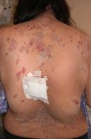 Erosions and healing areas on the back.
 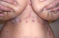 Healing areas on the chest and abdomen. Corticosteroids
have improved overall mortality, but now much of the mortality and
morbidity in these patients relates to the adverse effects of therapy.
Whether massive doses of steroids have any advantage over doses of 1
mg/kg/d is unclear.
Healing areas on the chest and abdomen. Corticosteroids
have improved overall mortality, but now much of the mortality and
morbidity in these patients relates to the adverse effects of therapy.
Whether massive doses of steroids have any advantage over doses of 1
mg/kg/d is unclear.
Immunosuppressive drugs are steroid sparing and should be considered early in the course of the disease. Epidermal growth factor may speed healing of localized lesions.[Many authorities now use rituximab as first- or second-line therapy. The antitumor necrosis factor drugs sulfasalazine and pentoxifylline have been reported as effective adjunctive treatments, reducing the serum level of tumor necrosis factor and resulting in rapid clinical improvement. Dapsone has been suggested as a steroid-sparing agent in the maintenance phase of pemphigus vulgaris treatment ; dapsone has also been suggested as a first-line agent.
Intravenous immunoglobulin therapy has been suggested as efficacious in pemphigus vulgaris treatment. Amagai et al reported on the successful use of intravenous immunoglobulin in pemphigus patients who did not fully respond to systemic steroids, and Asarch et al reported its use in pediatric patients.
Photodynamic therapy has been suggested as a possible adjunctive treatment for recalcitrant ulceration
Pemphigus is rare group of autoimmune diseases that Cause the formation of blisters. Pemphigus Herbal Treatment is aimed at reducing pain and preventing complications. Increasing number of sores and blisters are Symptoms of Pemphigus.
ReplyDeletepemphigus is treated with herbs. I had worse symptoms over my skin since 2013, I saw doctor Steven online and he gave me herbs remedies to use and now I have no symptoms and cured with my Herpes. I want you to contact Steven on WhatsApp to get information on this herbs ( +2348163807836 )
ReplyDelete