Today, the term scabies refers to the skin lesions produced by this mite. A readily treatable infestation, scabies remains common primarily because of diagnostic difficulty, inadequate treatment of patients and their contacts, and improper environmental control measures. Scabies is a great clinical imitator. Its spectrum of cutaneous manifestations and associated symptoms often results in delayed diagnosis. In fact, the phrase "7-year itch" was first used with reference to persistent, undiagnosed infestations with scabies.
Scabies is a worldwide public health problem, affecting persons of all ages, races, and socioeconomic groups. Overcrowding, delayed diagnosis and treatment, and poor public education contribute to the prevalence of scabies in both industrial and nonindustrial nations. Prevalence rates are higher in children and sexually active individuals than in other persons. Patients with poor sensory perception due to entities such as leprosy and persons with immunocompromise due to conditions such as status posttransplantation, HIV disease, and old age are at particular risk for the crusted variant. These populations present with clinically atypical lesions and often are misdiagnosed, thus delaying treatment and elevating the risk of local epidemics.
The
historical aspects of scabies infestations are quite reliable in
suggesting the diagnosis. Lesion distribution, intractable pruritus that
is worse at night, and similar symptoms in close contacts should
immediately rank scabies at the top of the clinical differential
diagnosis.
Lesion distribution differs in adults and children. Adults manifest lesions primarily on the flexor aspects of the wrists, the interdigital web spaces of the hands, the dorsal feet, axillae, elbows, waist, buttocks, and genitalia. Pruritic papules and vesicles on the scrotum and penis in men and areolae in women are highly characteristic.
Infants and small children may develop lesions diffusely, but unlike adults, lesions are common on the face, scalp, neck, palms, and soles. All cutaneous sites are susceptible in immunocompromised and elderly patients, who often have a history of a widespread, pruritic eczematous eruption.
Consider the diagnosis of scabies in any patient presenting with a recent onset of intense itching that is accentuated at night.
Physical
Clinical
findings include both primary and secondary lesions. Primary lesions
are the first manifestation of the infestation, and these typically
include small papules, vesicles, and burrows. Secondary lesions are the
result of rubbing and scratching, and they may be the only clinical
manifestation of the disease. If so, the diagnosis must be inferred by
the history, lesion distribution, and accompanying symptoms.
Burrows are a pathognomonic sign and represent the intraepidermal tunnel created by the moving female mite. They appear as serpiginous, grayish, threadlike elevations ranging from 2-10 millimeters long, as seen in the image below.
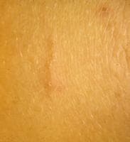 A typical linear burrow on the flexor forearm. Courtesy of Kenneth E. Greer, MD. They
are not readily apparent and must be actively sought. A black dot may
be seen at one end of the burrow, indicating the presence of a mite.
High-yield locations for burrows include the webbed spaces of the
fingers, flexor surfaces of the wrists, elbows, axillae, belt line,
feet, scrotum in men, and areolae in women. In infants, burrows are
commonly located on the palms and soles, as in the image below. The
actual mites are microscopic and cannot be visualized with the unaided
human eye.
A typical linear burrow on the flexor forearm. Courtesy of Kenneth E. Greer, MD. They
are not readily apparent and must be actively sought. A black dot may
be seen at one end of the burrow, indicating the presence of a mite.
High-yield locations for burrows include the webbed spaces of the
fingers, flexor surfaces of the wrists, elbows, axillae, belt line,
feet, scrotum in men, and areolae in women. In infants, burrows are
commonly located on the palms and soles, as in the image below. The
actual mites are microscopic and cannot be visualized with the unaided
human eye.
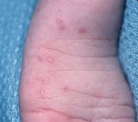 A
subtle linear burrow accompanied by erythematous papules on the sole of
the foot in a child with scabies. Courtesy of Kenneth E. Greer, MD. One-
to 3-mm erythematous papules and vesicles are seen in typical
distributions in adults. The vesicles are discrete lesions filled with
clear fluid, although the fluid may appear cloudy if the vesicle is more
than a few days old, as in the image below.
A
subtle linear burrow accompanied by erythematous papules on the sole of
the foot in a child with scabies. Courtesy of Kenneth E. Greer, MD. One-
to 3-mm erythematous papules and vesicles are seen in typical
distributions in adults. The vesicles are discrete lesions filled with
clear fluid, although the fluid may appear cloudy if the vesicle is more
than a few days old, as in the image below.
 Erythematous papules and papulovesicles on the flexor wrist. Courtesy of Kenneth E. Greer, MD. Papules
rarely contain mites and most likely represent a hypersensitivity
reaction. Papules are common on the shaft of the penis in men and on the
areolae in women, as in the images below.
Erythematous papules and papulovesicles on the flexor wrist. Courtesy of Kenneth E. Greer, MD. Papules
rarely contain mites and most likely represent a hypersensitivity
reaction. Papules are common on the shaft of the penis in men and on the
areolae in women, as in the images below.
 Scabies on the penile shaft and glans. Courtesy of William D. James, MD.
 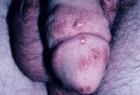 Scabietic papules on the penile shaft and scrotum. Courtesy of Kenneth E. Greer, MD. Unlike adults, who rarely present with facial and neck involvement, this presentation is fairly typical in children.
In very young children and infants, a widespread eczematous eruption primarily on the trunk is common, as in the image below.
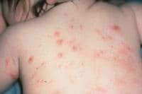 Widespread eruption on the back of an infant with scabies. Courtesy of Kenneth E. Greer, MD. In
neonates unable to scratch, pinkish-brown nodules may develop and range
in size from 2-20 mm, as demonstrated in the images below. Mites are
rarely found within the nodules. Infants may have 1 to 3-mm papules,
vesicles, and pustules on the palms and soles.
Widespread eruption on the back of an infant with scabies. Courtesy of Kenneth E. Greer, MD. In
neonates unable to scratch, pinkish-brown nodules may develop and range
in size from 2-20 mm, as demonstrated in the images below. Mites are
rarely found within the nodules. Infants may have 1 to 3-mm papules,
vesicles, and pustules on the palms and soles.
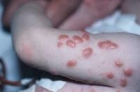 Nodular scabies in an infant. Courtesy of Kenneth E. Greer, MD.
 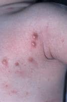 Nodular scabies. Courtesy of Kenneth E. Greer, MD. Crusted
scabies, previously referred to as Norwegian scabies, manifests with
marked thickening and crusting of the skin. Lesions are often
hyperkeratotic, crusted, and cover large areas. Marked scaling is
common, and pruritus may be minimal or absent. Nail dystrophy and scalp
lesions may be prominent. The hands and arms are usual locations, but
all sites are vulnerable, as demonstrated in the images below. Mites can
number in the thousands to millions in this form. Predominantly
affected are those with immunosuppression, neurological disorders, or
institutionalization. Possible risk factors for profound infestation in
these specific populations include an inability to mount an immune
response, perceive pruritus, and/or physically scratch the skin (a
mechanism to rid the body of mites).
Nodular scabies. Courtesy of Kenneth E. Greer, MD. Crusted
scabies, previously referred to as Norwegian scabies, manifests with
marked thickening and crusting of the skin. Lesions are often
hyperkeratotic, crusted, and cover large areas. Marked scaling is
common, and pruritus may be minimal or absent. Nail dystrophy and scalp
lesions may be prominent. The hands and arms are usual locations, but
all sites are vulnerable, as demonstrated in the images below. Mites can
number in the thousands to millions in this form. Predominantly
affected are those with immunosuppression, neurological disorders, or
institutionalization. Possible risk factors for profound infestation in
these specific populations include an inability to mount an immune
response, perceive pruritus, and/or physically scratch the skin (a
mechanism to rid the body of mites).
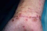
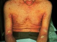 Crusted scabies. Courtesy of William D. James, MD.
 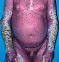 Crusted scabies. Courtesy of Kenneth E. Greer, MD. Nodular
scabies occurs in 7-10% of patients with scabies, particularly young
children as noted above. Pink, tan, brown, or red nodules may be
present, ranging from 2-20 millimeters in diameter.
Crusted scabies. Courtesy of Kenneth E. Greer, MD. Nodular
scabies occurs in 7-10% of patients with scabies, particularly young
children as noted above. Pink, tan, brown, or red nodules may be
present, ranging from 2-20 millimeters in diameter.
Characteristic findings include excoriations, widespread eczema, honey-colored crusting, postinflammatory hyperpigmentation, erythroderma, prurigo nodules, and frank pyoderma.[3, 4]
Primary scabies lesions
The distribution is highly characteristic in typical cases.Burrows are a pathognomonic sign and represent the intraepidermal tunnel created by the moving female mite. They appear as serpiginous, grayish, threadlike elevations ranging from 2-10 millimeters long, as seen in the image below.
A typical linear burrow on the flexor forearm. Courtesy of Kenneth E. Greer, MD. They
are not readily apparent and must be actively sought. A black dot may
be seen at one end of the burrow, indicating the presence of a mite.
High-yield locations for burrows include the webbed spaces of the
fingers, flexor surfaces of the wrists, elbows, axillae, belt line,
feet, scrotum in men, and areolae in women. In infants, burrows are
commonly located on the palms and soles, as in the image below. The
actual mites are microscopic and cannot be visualized with the unaided
human eye.A
subtle linear burrow accompanied by erythematous papules on the sole of
the foot in a child with scabies. Courtesy of Kenneth E. Greer, MD. One-
to 3-mm erythematous papules and vesicles are seen in typical
distributions in adults. The vesicles are discrete lesions filled with
clear fluid, although the fluid may appear cloudy if the vesicle is more
than a few days old, as in the image below.Erythematous papules and papulovesicles on the flexor wrist. Courtesy of Kenneth E. Greer, MD. Papules
rarely contain mites and most likely represent a hypersensitivity
reaction. Papules are common on the shaft of the penis in men and on the
areolae in women, as in the images below.Scabies on the penile shaft and glans. Courtesy of William D. James, MD. Scabietic papules on the penile shaft and scrotum. Courtesy of Kenneth E. Greer, MD. Unlike adults, who rarely present with facial and neck involvement, this presentation is fairly typical in children.In very young children and infants, a widespread eczematous eruption primarily on the trunk is common, as in the image below.
Widespread eruption on the back of an infant with scabies. Courtesy of Kenneth E. Greer, MD. In
neonates unable to scratch, pinkish-brown nodules may develop and range
in size from 2-20 mm, as demonstrated in the images below. Mites are
rarely found within the nodules. Infants may have 1 to 3-mm papules,
vesicles, and pustules on the palms and soles.Nodular scabies in an infant. Courtesy of Kenneth E. Greer, MD. Nodular scabies. Courtesy of Kenneth E. Greer, MD. Crusted
scabies, previously referred to as Norwegian scabies, manifests with
marked thickening and crusting of the skin. Lesions are often
hyperkeratotic, crusted, and cover large areas. Marked scaling is
common, and pruritus may be minimal or absent. Nail dystrophy and scalp
lesions may be prominent. The hands and arms are usual locations, but
all sites are vulnerable, as demonstrated in the images below. Mites can
number in the thousands to millions in this form. Predominantly
affected are those with immunosuppression, neurological disorders, or
institutionalization. Possible risk factors for profound infestation in
these specific populations include an inability to mount an immune
response, perceive pruritus, and/or physically scratch the skin (a
mechanism to rid the body of mites).Crusted scabies. Courtesy of William D. James, MD. Crusted scabies. Courtesy of Kenneth E. Greer, MD. Nodular
scabies occurs in 7-10% of patients with scabies, particularly young
children as noted above. Pink, tan, brown, or red nodules may be
present, ranging from 2-20 millimeters in diameter. Secondary scabies lesions
These are the result of scratching, secondary infection, and/or the host immune response against the mites and their products.Characteristic findings include excoriations, widespread eczema, honey-colored crusting, postinflammatory hyperpigmentation, erythroderma, prurigo nodules, and frank pyoderma.[3, 4]
Causes
Human scabies is caused by the host-specific mite, S scabiei var hominis, an obligate human parasite. It is a member of the class Arachnida, subclass Acari, order Astigmata, and family Sarcoptidae.
Human infestation with S scabiei varieties of animal origin can occur. Both domestic and wild animals worldwide are susceptible to infestation with S scabiei, and the resultant disease is referred to as sarcoptic mange. Mange due to S scabiei varieties other than hominis has been reported in dogs, pigs, horses, camels, black bears, monkeys, dingoes, and wild fox, among others. Although reports have described transfer to humans from animals, experimental studies have demonstrated limited cross-infectivity between different host species. Further, genotyping studies have revealed that the Sarcoptes mites segregate into separate host-associated populations, thus limiting the transmission across host species.
In the rare instance of transmission of nonhuman scabies from animals to humans, the clinical manifestations differ in many respects. The incubation period is shorter, the symptoms are transient, the infestation is self-limiting, no burrows are formed, and the distribution is atypical compared with infestation caused by S scabiei var hominis. Contacts of patients with scabies contracted from an animal source require no treatment.
Treatment for scabies includes administration of a scabicidal agent, an antipruritic agent such as a sedating antihistamine, and an appropriate antimicrobial agent if secondarily infected.
Provision of detailed verbal and written instructions is critical for compliance and complete eradication of scabies.
All family members and close contacts must be evaluated and treated for scabies, even if they do not have symptoms. Pets do not require treatment. All carpets and upholstered furniture should be vacuumed and vacuum bags immediately discarded.
Instruct patients to launder clothing, bed linens, and towels used within the last week in hot water the day after treatment is initiated and again in 1 week. Items that cannot be washed may be professionally dry cleaned or sealed in plastic bags for 1 week.
Patients with crusted scabies or their caregivers should be instructed to remove excess scale to allow penetration of the topical scabicidal agent and decrease the burden of infestation. This can be achieved with warm water soaks followed by application of a keratolytic agent such as 5% salicylic acid in petrolatum or Lac-Hydrin cream. (Salicylic acid should be avoided if large body surface areas are involved because of the potential risk of salicylate poisoning.) The scales are then mechanically debrided with a tongue depressor or similar nonsharp device.
No comments:
Post a Comment