In 1909, Hyde and Montgomery. first
described prurigo nodularis as pruritic nodules on the extensor
surfaces of the lower extremities in middle-aged women. Prurigo
nodularis can be a bothersome-to-debilitating disease, usually seen as
multiple, intensely pruritic, excoriated nodules erupting on the
extensor surfaces of the limbs secondary to itching or rubbing. In
current practice, prurigo nodularis is still a condition of unknown
etiology. Many conditions have been reported to induce prurigo
nodularis, from internal malignancy to renal failure to psychiatric
conditions.
Prurigo
nodularis patients are most often middle-aged to elderly. Patients with
prurigo nodularis invariably complain of a long-standing history of
severe, unremitting pruritus. Patients can point out specific sites
where they began feeling itchy and where dark-colored nodules formed
soon after. Mature nodules rarely increase or decrease in size;
spontaneous resolution is even more rare. Prurigo nodularis is usually
bilaterally symmetric, with nodules that are either stable or increasing
in number.
The patient's medical history may be significant for several conditions, as follows:
The patient's medical history may be significant for several conditions, as follows:
- Hepatic or renal dysfunction
- Local trauma or insult to the skin
- Infection
- HIV/immunodeficiency[2]
- Anxiety or other psychiatric condition
Physical
Prurigo
nodularis nodules or papules are 3-20 mm in diameter; they are
discrete, scaly, generally symmetric, hyperpigmented or purpuric, and
firm. Nodules and papules occur on the extensor surfaces of the arms,
the legs, and sometimes the trunk.
Prurigo nodularis lesions may show signs of excoriation with flat, umbilicated, or crusted top. Lesions may number from 1-2 to hundreds. The nodule pattern may be follicular. Upon entering the examination room and while patients' describe the locations of the lesions, patients may scratch or rub the lesions rather than pointing to them. Many prurigo nodularis patients appear very anxious, worried, or even obsessed with the nodules. Note the images below.
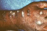 Prurigo nodularis. Courtesy of Jeffrey Meffert, MD.
 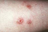 Prurigo nodularis. Courtesy of Jeffrey Meffert, MD.
Prurigo nodularis lesions may show signs of excoriation with flat, umbilicated, or crusted top. Lesions may number from 1-2 to hundreds. The nodule pattern may be follicular. Upon entering the examination room and while patients' describe the locations of the lesions, patients may scratch or rub the lesions rather than pointing to them. Many prurigo nodularis patients appear very anxious, worried, or even obsessed with the nodules. Note the images below.
Prurigo nodularis. Courtesy of Jeffrey Meffert, MD. Prurigo nodularis. Courtesy of Jeffrey Meffert, MD. Causes
The
cause of prurigo nodularis is still unknown. Many associated conditions
are known, but their roles as coexisting or preexisting conditions have
not been established in causing prurigo nodularis. Notable changes in
papules and nodules are increased in certain inflammatory cell types,
inflammatory products, and neural hyperplasia.
Mast cells and neutrophils are seen in higher-than-normal levels in prurigo nodularis; however, their degranulation products are not increased. Eosinophils are not seen in higher numbers; however, the protein granule products (ie, major basic protein, eosinophil cationic protein, eosinophil-derived neurotoxin) are seen in significantly higher levels.
Papillary dermal nerves and Merkel cells are sensory nerves found in the dermis and the epidermis, respectively. They are both found in increased numbers in prurigo nodularis. These are neural receptors that sense touch, temperature, pain, and itch. These increases in sensory nerves are not seen in lichen simplex chronicus, another pruritic disease that causes epidermal hyperplasia but in a plaquelike morphology.
Calcitonin gene–related peptide and substance P immunoreactive nerves are markedly increased in prurigo nodularis skin compared with normal skin. These neuropeptides may mediate the cutaneous neurogenic inflammation and pruritus in prurigo nodularis. In addition, the capsaicin-binding nonselective cation channel known as vanilloid receptor subtype 1 has highly increased expression in epidermal keratinocytes and nerve fibers in prurigo nodularis lesions, but these can be normalized with capsaicin application.
Hepatitis C, mycobacteria, Helicobacter pylori, Strongyloides stercoralis, and HIV have been reported as infectious etiologies of prurigo nodularis or as associated with prurigo nodularis in case reports or from single-center studies.
Interleukin 31, a T-cell–derived cytokine that causes severe pruritus and dermatitis in transgenic mice, is elevated in individuals with prurigo nodularis.Interleukin 31 expression in atopic individuals is also rapidly induced by staphylococcal superantigen; however, the link between these findings has not been extensively researched.
Current available treatments of prurigo nodularis have had mild-to-moderate success at best. Often, combinations of several medications or physical modalities may be used in an attempt to control this process.
Topical, oral, and intralesional corticosteroids have all been used in prurigo nodularis in attempts to decrease inflammation and sense of itching and to soften and smooth out firm nodules. The improvement with corticosteroids is variable, and corticosteroids are sometimes not helpful. Intralesional corticosteroid (usually triamcinolone acetonide) treatment is commonly used in resistant cases of limited extent. Triamcinolone acetonide concentrations as low as 2.5 mg/mL may be effective, although more scarred pruriginous lesions may require higher concentrations. A total dose of 20 mg (8 mL of 2.5 mg/mL) for adults every 3-4 weeks is safe for patients without diabetes mellitus or hypertension. An 8-am serum cortisol test can be performed if concerns exist about adrenal suppression.
Menthol, phenol, pramoxine, capsaicin cream, vitamin D-3 ointment, and topical anesthetics are some other topical agents used to reduce pruritus. Treatment with DuoDerm or other occlusive therapies has been suggested to flatten lesions while at the same time preventing patients from directly scratching nodules.
UV light treatment using UV-B or UV-A plus psoralen may be beneficial for severe pruritus. Consider the adverse effects of prolonged UV exposure before such treatment. Monochromatic 308-nm light therapy may be helpful for recalcitrant lesions, although this modality may be more useful in atopic dermatitis. UV-A1 has also been reported to benefit lichen simplex chronicus and prurigo nodularis.
Antihistamines, anxiolytics, opiate receptor antagonists, and (most recently) thalidomide are oral medications other than steroids used for prurigo nodularis. Thalidomid. has been shown to aid in several severe dermatoses, including prurigo nodularis with or without associated HIV disease.Severe teratogenic effects are well known and documented, and all women of childbearing age should be on adequate birth control methods. Patients taking thalidomide have an increased risk of peripheral neuropathy.
For steroid unresponsive patients or those with lesions on thin skin, a few case reports and small studies have shown efficacy of the topical immunomodulators tacrolimus and pimecrolimus.
Anecdotally, gabapentin has been reported to benefit prurigo nodularis. Sedation is the main problem with this generic medication.
Habit reversal therapy for the itch-scratch cycle associated with prurigo nodularis may be helpful and can be administered by dermatology nurses trained in this therapy.
No comments:
Post a Comment