Impetigo is an acute, highly
contagious gram-positive bacterial infection of the superficial layers
of the epidermis. Impetigo occurs most commonly in children, especially
those who live in hot, humid climates. The name is believed to be
derived from the Latin impetere (to assail).
Impetigo occurs in 2 forms: bullous and nonbullous, as shown in the photographs below. Nonbullous impetigo is the more common form, constituting approximately 70% of impetigo cases. It tends to affect skin on the face or extremities that has been disrupted by bites, cuts, abrasions, other trauma, or diseases such as varicella.[2]
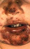 Nonbullous impetigo with vesicles, pustules, and sharply demarcated regions of honey-colored crusts.
 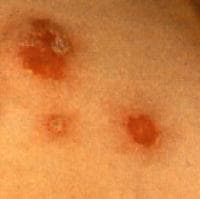 Bullous impetigo with circumscribed lesions with a thin collarette of scale. Nonbullous
impetigo, also known as impetigo contagiosa, is the most common skin
infection in children, accounting for approximately 10% of all cutaneous
problems in pediatric clinics. It is more contagious than the bullous
type. Common
impetigo is the term applied when the infection occurs in preexisting
wounds. Impetigo as a secondary infection of preexisting skin disease or
traumatized skin has also been referred to as impetiginous dermatitis.
Bullous impetigo with circumscribed lesions with a thin collarette of scale. Nonbullous
impetigo, also known as impetigo contagiosa, is the most common skin
infection in children, accounting for approximately 10% of all cutaneous
problems in pediatric clinics. It is more contagious than the bullous
type. Common
impetigo is the term applied when the infection occurs in preexisting
wounds. Impetigo as a secondary infection of preexisting skin disease or
traumatized skin has also been referred to as impetiginous dermatitis.
Nonbullous impetigo is caused by Staphylococcus aureus,group A beta hemolytic streptococci (GABHS, also known as Streptococcus pyogenes), or a combination of both. Most infections begin as a streptococcal infection, but staphylococci replace the streptococci over time.
Methicillin-resistant S aureus (MRSA), which can be hospital or community acquired, is an increasingly common cause of impetigo ; this pathogen is observed more often with the nonbullous form of impetigo than the bullous form. Over the last decade, an increasing number of community-acquired MRSA and gentamicin-resistant S aureus strains have been reported as a cause of impetigo.
Bullous impetigo may affect intact skin and is caused almost exclusively by S aureus. Bullous impetigo is a toxin-mediated erythroderma in which the epidermal layer of the skin sloughs, resulting in large areas of skin loss. Ecthyma is a deeper, ulcerated infection, often occurring with lymphadenitis, that may be a complication of impetigo.
Impetigo seldom progresses to systemic infection, although poststreptococcal glomerulonephritis is a rare complication with GABHS infection only. Certain serotypes of GABHS (eg, types 49, 55, 57, 59) are associated with impetigo and acute glomerulonephritis.
Impetigo can also present as folliculitis, which is considered to be impetigo of the hair follicles caused by S aureus. Chronic recalcitrant impetigo/folliculitis can result in sycosis barbae (similar to lupoid sycosis) with scarring and a presentation similar to that of discoid lupus. Tinea may also cause this presentation.
Diagnosis of impetigo is usually based solely on the history and clinical appearance (see Presentation and Workup). Treatment typically involves local wound care, along with antibiotic therapy, either topical or systemic plus topical (see Treatment and Medication).
Impetigo occurs in 2 forms: bullous and nonbullous, as shown in the photographs below. Nonbullous impetigo is the more common form, constituting approximately 70% of impetigo cases. It tends to affect skin on the face or extremities that has been disrupted by bites, cuts, abrasions, other trauma, or diseases such as varicella.[2]
Nonbullous impetigo with vesicles, pustules, and sharply demarcated regions of honey-colored crusts. Bullous impetigo with circumscribed lesions with a thin collarette of scale. Nonbullous
impetigo, also known as impetigo contagiosa, is the most common skin
infection in children, accounting for approximately 10% of all cutaneous
problems in pediatric clinics. It is more contagious than the bullous
type. Common
impetigo is the term applied when the infection occurs in preexisting
wounds. Impetigo as a secondary infection of preexisting skin disease or
traumatized skin has also been referred to as impetiginous dermatitis. Nonbullous impetigo is caused by Staphylococcus aureus,group A beta hemolytic streptococci (GABHS, also known as Streptococcus pyogenes), or a combination of both. Most infections begin as a streptococcal infection, but staphylococci replace the streptococci over time.
Methicillin-resistant S aureus (MRSA), which can be hospital or community acquired, is an increasingly common cause of impetigo ; this pathogen is observed more often with the nonbullous form of impetigo than the bullous form. Over the last decade, an increasing number of community-acquired MRSA and gentamicin-resistant S aureus strains have been reported as a cause of impetigo.
Bullous impetigo may affect intact skin and is caused almost exclusively by S aureus. Bullous impetigo is a toxin-mediated erythroderma in which the epidermal layer of the skin sloughs, resulting in large areas of skin loss. Ecthyma is a deeper, ulcerated infection, often occurring with lymphadenitis, that may be a complication of impetigo.
Impetigo seldom progresses to systemic infection, although poststreptococcal glomerulonephritis is a rare complication with GABHS infection only. Certain serotypes of GABHS (eg, types 49, 55, 57, 59) are associated with impetigo and acute glomerulonephritis.
Impetigo can also present as folliculitis, which is considered to be impetigo of the hair follicles caused by S aureus. Chronic recalcitrant impetigo/folliculitis can result in sycosis barbae (similar to lupoid sycosis) with scarring and a presentation similar to that of discoid lupus. Tinea may also cause this presentation.
Diagnosis of impetigo is usually based solely on the history and clinical appearance (see Presentation and Workup). Treatment typically involves local wound care, along with antibiotic therapy, either topical or systemic plus topical (see Treatment and Medication).
Nonbullous impetigo begins with a
single erythematous macule that rapidly evolves into a vesicle or
pustule and ruptures; the released serous contents then dry, leaving a
crusted, honey-colored exudate over the erosion. Rapid spread follows by
contiguous extension or to distal areas through inoculation of other
wounds from scratching.
Skin on any part of the body can be involved, but the face and extremities are affected most commonly. Lesions are usually asymptomatic, with occasional pruritus. Little or no surrounding erythema or edema is present. Regional adenopathy is common.
Patients with impetigo may report a history of minor trauma, insect bites, scabies, herpes simplex, varicella, or eczema at the site of infection. Any history of preexisting skin disease should raise the clinician's index of suspicion.
Bullous impetigo usually consists of small or large, superficial, fragile bullae. Often, these quickly appear, spontaneously rupture, and drain so that only the remnants, or collarettes, are seen at the time of presentation. The lesions usually spread locally in the face, trunk, extremities, buttocks, or perineal regions and may reach distal areas through direct autoinoculation.
Lesions typically appear on intact skin but may secondarily invade preexisting lesions (eg, eczema) to cause generalized lesions. There is minimal or no surrounding erythema and no regional lymphadenopathy.
Individuals with impetigo frequently recall exposure to a person who is a known carrier of S aureus or streptococcal organisms, has a pyoderma, or has a skin condition (eg, atopic dermatitis) that predisposes that individual to be an S aureus or streptococcal carrier. Clusters in families and outbreaks in institutions are occasionally reported.
Hot humid weather, participation in contact sports, crowded living conditions, poor personal hygiene, or an unhygienic work environment encourages contamination of the skin by pathogenic bacteria that can cause impetigo.
Conditions such as HIV infection, posttransplantation, diabetes mellitus, hemodialysis, chemotherapy, radiation therapy, or systemic corticosteroid treatment increase susceptibility.
Primary selective immunoglobulin M (IgM) deficiency has been linked to recurrent impetigo in patients with negative S aureus carrier status and no predisposing factors, such as a preexisting dermatosis.[26] Frequent associations of immunoglobulin A (IgA), IgM, and immunoglobulin G (IgG) deficiencies have also been reported.
The following symptoms usually are absent in impetigo contagiosa but may be present in bullous impetigo:
- Fever
- Diarrhea
- Generalized weaknessTreatment of impetigo typically involves local wound care along with antibiotic therapy. Antibiotic therapy for impetigo may be with a topical agent alone or a combination of systemic and topical agents.
Gentle cleansing, removal of the honey-colored crusts of nonbullous impetigo using antibacterial soap and a washcloth, and frequent application of wet dressings to areas affected by lesions are recommended. Good hygiene with antibacterial washes, such as chlorhexidine, may prevent the transmission of impetigo and prevent recurrences, but the efficacy of this has not been proven.
For antibiotic therapy, the chosen agent must provide coverage against both Staphylococcus aureus and Streptococcus pyogenes. Community-acquired methicillin-resistant S aureus (MRSA) infection most commonly manifests as folliculitis or abscess, rather than impetigo; thus, beta-lactam drugs remain an appropriate initial empiric choice.
Topical mupirocin is adequate treatment for single lesions of nonbullous impetigo or small areas of involvement. It is applied to the affected area 2 to 3 times daily. A 7-day course is usually standard, although few large studies have been performed to verify this as the most effective approach. Systemic antibiotics are indicated for extensive involvement or for bullous impetigo.
In patients with bullous impetigo who present to the emergency department with large areas of involvement resulting in denuded skin from ruptured bullae, management also includes intravenous fluid resuscitation. Fluid is given at a volume and rate similar to standard volume replacement for burns.
Inpatient care is required for patients with impetigo who have widespread disease or for infants at risk of sepsis and/or dehydration due to skin loss. If inpatient care is warranted in the child with untreated impetigo, contact isolation is recommended.
A clinical guideline summary from the Infectious Diseases Society of America that includes recommendations on impetigo..
your tips are very useful. thanks for posting.
ReplyDeleteskin specialist